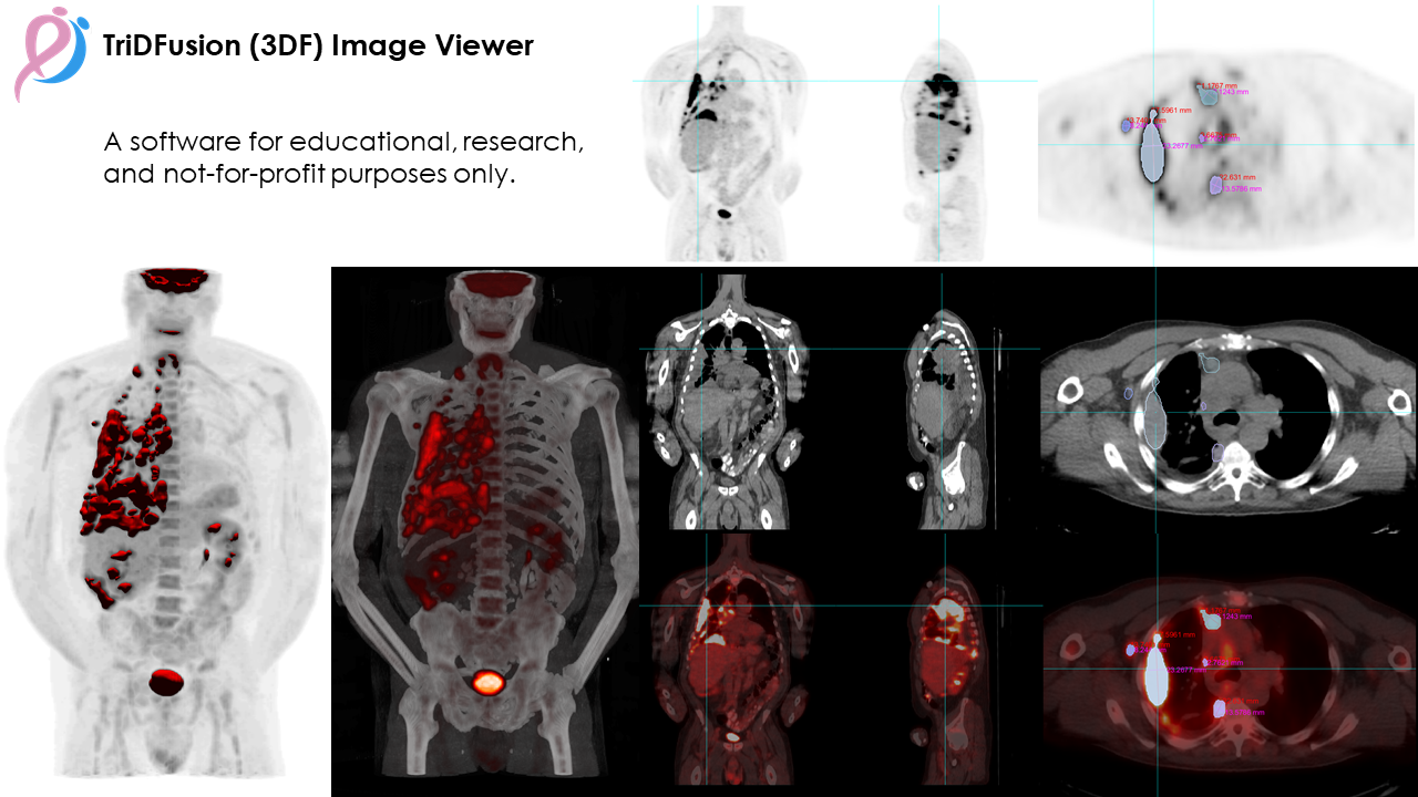
The TriDFusion (3DF) image Viewer is a powerful research tool designed specifically for physicists. This advanced viewer is capable of handling multiple modalities and dimensions of DICOM images, with dynamic memory allocation to ensure smooth performance. It can address common issues such as image orientation, slice separation, and miss-registration, and can even remove and set hot pixels, such as those found in Whole-Body SPECT planar bladder images.
One of the key features of the 3DF Image Viewer is its mask and image segmentation tool, which works seamlessly with any modality. This allows researchers to easily isolate specific areas of interest and generate high-quality images for further analysis. These images can be exported as a new DICOM series or in .stl format for 3D printing.
In addition, the 3DF Image Viewer offers powerful fusion capabilities, allowing researchers to combine MIP with isosurface imaging for improved lesion detection and characterization in 18F-FDG PET/CT scans. Furthermore, the addition of volume rendering with customized color and alpha maps can provide even more information about the intensity of uptake in lesions.
For tumor quantification, the 3DF Image Viewer offers a 3D threshold-based isosurface that can be easily exported as an stl file or as a new DICOM 3D mask. With its advanced features and robust capabilities, the TriDFusion (3DF) Image Viewer is an essential tool for physicists seeking to push the boundaries of medical imaging research.
The TriDFusion (3DF) image viewer is published in the EJNMMI Physics journal and is accessible online here.
If you use TriDFusion (3DF) please cite it:
Lafontaine D, Schmidtlein CR, Kirov A, Reddy RP, Krebs S, Schöder H, Humm JL. TriDFusion (3DF) image viewer. EJNMMI Phys. 2022 Oct 18;9(1):72. doi: 10.1186/s40658-022-00501-y. PMID: 36258098; PMCID: PMC9579267.
| Medical Imaging Modalities | Import / Export File Formats |
|---|---|
| Positron Emission Tomography PET-CT (PT) | DICOM using custom/vendor dictionaries |
| Gamma Camera, Nuclear Medicine (NM) | Raw data from nuclear imaging devices |
| Computed Tomography (CT) | DICOM-RT structure (contours) |
| Digital Radiography (CR, DX) | CERR planC, dose volumes and constraints |
| Digital Angiography (XA) | Comma Separated Values (.csv) |
| Magnetic Resonance (MR) | Standard Triangle Language (.stl) |
| Secondary Pictures and Scanned Images (SC) | Audio Video Interleave (.avi) |
| Mammography (MG) | Moving Pictures Expert Group 4 (.mp4) |
| Ultrasonography (US) | Graphics Interchange Format (.gif) |
| Digital Imaging and Communications (.dcm) | |
| Joint Photographic Experts Group (.jpg) | |
| Bitmap (.bmp) | |
| Portable Network Graphics (.png) | |
| Neuroimaging Informatics Technology Initiative (.nii) | |
| Nearly Raw Raster Data (.nrrd) |
Main features:
- Multi-modality Image Viewer
- Total Tumor Burden Determination
- 3D Visualization
- 3D Printing
- Image Multi-Fusion
- Image Convolution
- Image Registration
- Image Resampling
- Image Re-Orientation
- Image Arithmetic and Post Filtering
- Image Editing
- Image Mask
- Image Constraint
- Lung Segmentation
- Edge Detection
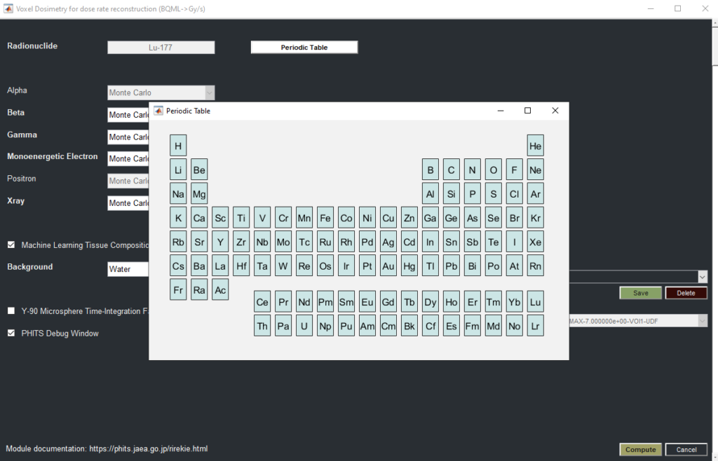
- Voxel Dosimetry
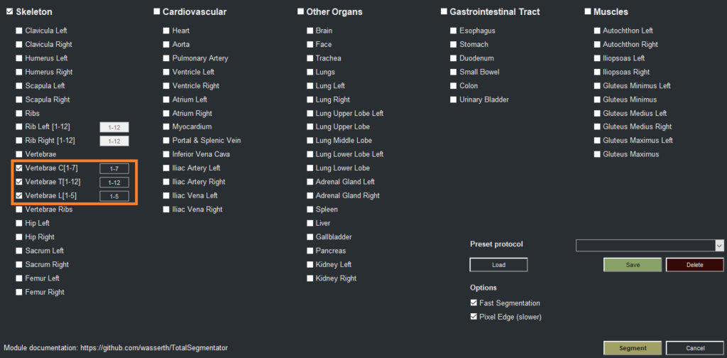
- Machine Learning Segmentation
- Machine Learning 3D Lung Shunt & Lung Dose
- Machine Learning 3D Lung Lobe Quantification
- Machine Learning Y90 Dosimetry
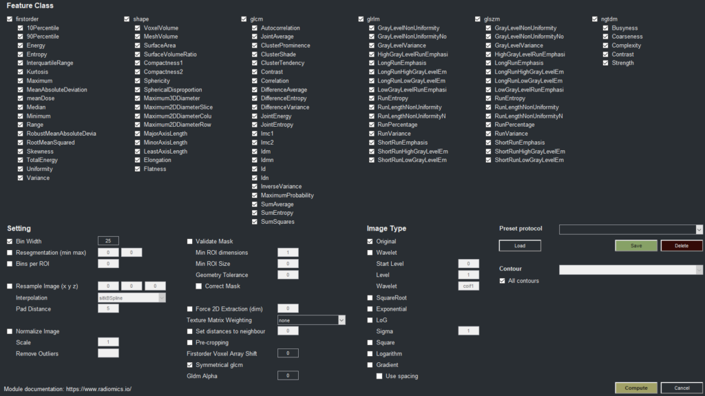
- Radiomics
Visit GitHub to download the source code.
Matlab compiled version can be downloaded here.
Examples:
Publications:

Lafontaine D, Schmidtlein CR, Kirov A, Reddy RP, Krebs S, Schöder H, Humm JL. TriDFusion (3DF) image viewer. EJNMMI Phys. 2022 Oct 18;9(1):72. doi: 10.1186/s40658-022-00501-y. PMID: 36258098; PMCID: PMC9579267.

Daniel Lafontaine, Charles Schmidtlein, Adam Kesner, Assen Kirov, Joseph O’Donoghue and John Humm
Journal of Nuclear Medicine June 2023, 64 (supplement 1) P495
Krebs, Simone; Lafontaine, D; Bodei, L; Mayerhoefer, ME; Humm, J; Schoder, H (2023). Deakin University. Journal contribution. https://hdl.handle.net/10779/DRO/DU:24945177.v1
Project Manager and Programmer:
Daniel Lafontaine*
Contributors:
C. Ross Schmidtlein*, Brad Beattie*, Assen Kirov*, Aditya Apte*, Adam Kesner*, Joseph O’Donoghue*, Alexandre Franca Velo*, Simone Krebs**, Lukas Carter*
Memorial Sloan Kettering
*Department of Medical Physics, New York, NY, USA
**Department of Radiology, New York, NY, USA
← Pojects